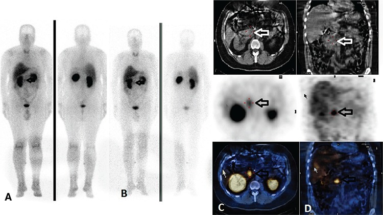
Figure 2. Iodine-123 metaiyodobenzilguanidin scintigraphy findings of case 2. Anterior-posterior whole body images show focal tracer uptake superior to the kidneys bilaterally (A) at 4, and (B) 24 hours, before surgery (arrows), (C) axial (D) coronal computed tomography (CT), single photon emission computerized tomography (SPECT), and SPECT/CT images reveal that those radiotracer accumulations are localized to the adrenal glands bilaterally (arrows). Histopathologic examination of the masses were reported as pheochromocytoma after surgery.

