Figure 5.
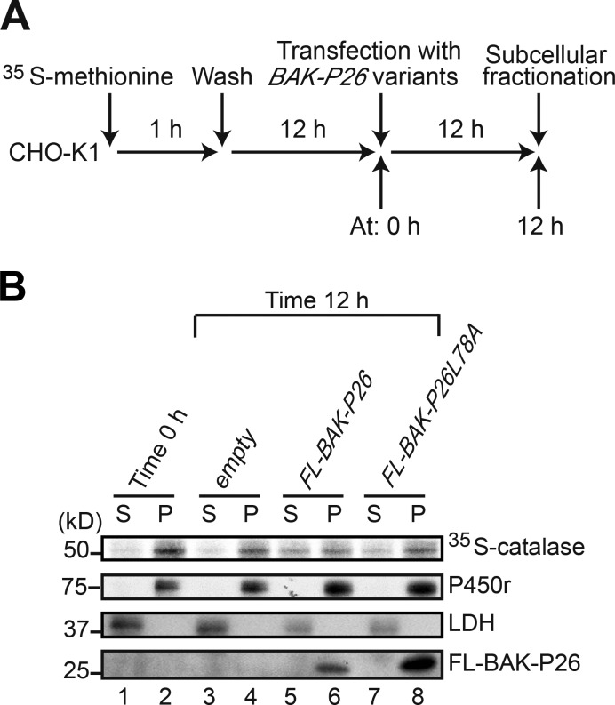
Peroxisome-targeted BAK releases catalase from peroxisomes to cytosol. (A) Time flow of pulse-chase experimentation of catalase translocation. (B) Cells at 0 h (before transfection) and 12 h after transfection with the indicated plasmids (time 12 h) were fractionated into cytosol (S) and organelle (P) fractions. Equal aliquots of cytosol and organelle fractions were solubilized, and catalase was immunoprecipitated with anticatalase antibody. [35S]methionine– and [35S]cysteine–labeled catalase was detected by a Fujix FLA-5000 autoimaging analyzer. As marker proteins for cytosol and the ER, lactate dehydrogenase (LDH) and P450 reductase (P450r) in S and P fractions were detected with the respective antibodies. BAK-P26 variants were detected with anti-Flag antibody.

