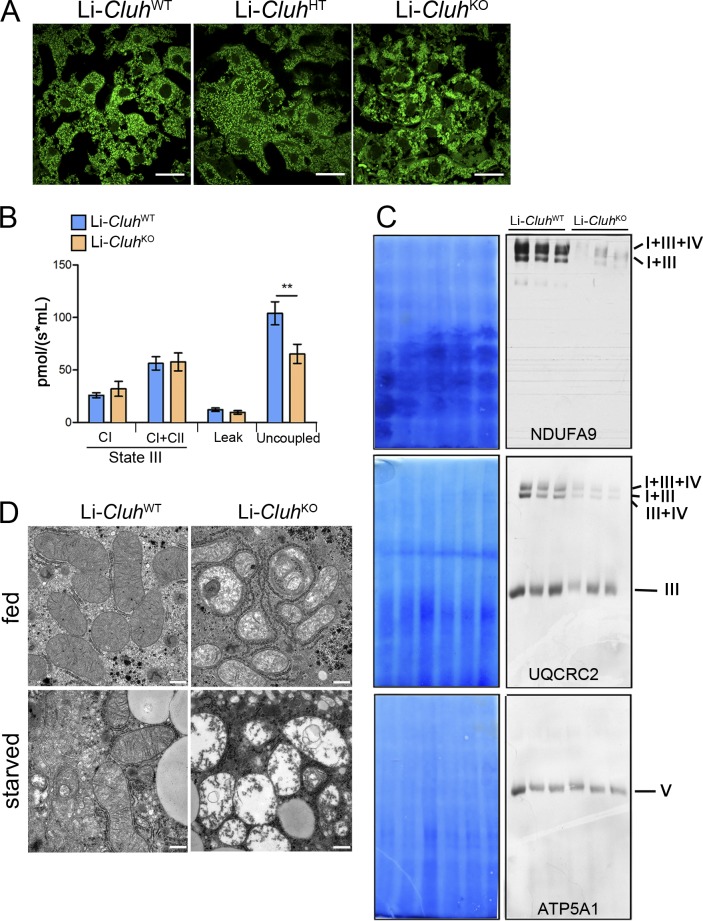
Figure 5.
Liver-specific Cluh deletion affects mitochondrial distribution and structure, assembled respiratory supercomplexes, and respiratory capacity. (A) Representative confocal images of livers of 8-wk-old mice of the indicated genotypes. To analyze mitochondrial morphology, mice were crossed with a stop-mito-YFP reporter line activated by Cre recombination. n = 4. Bars, 5 µm. (B) Oxygen consumption of mitochondria isolated from livers of 8-wk-old mice. State III respiration was measured in the presence of pyruvate, malate, glutamate, and ADP (complex I [CI]), followed by addition of succinate (complex I + complex II [CI + CII]). The proton leak was measured after addition of oligomycin, whereas maximal respiration was assessed by CCCP titration. n = 5. Graph shows means ± SEM. **, P ≤ 0.01 (two-tailed t test). (C) Blue-native–PAGE of digitonin-solubilized mitochondria isolated from the liver of mice with the indicated genotypes at 8 wk of age. Corresponding Coomassie stainings are shown. Supercomplexes were detected using antibodies against NDUFA9 (complex I) and UQCRC2 (complex III). ATP5A1 was used to detect complex V. (D) Representative electron micrographs showing mitochondrial structures in the livers of 8-wk-old mice either fed ad libitum or starved for 24 h. n = 3. Bars, 0.5 µm.

