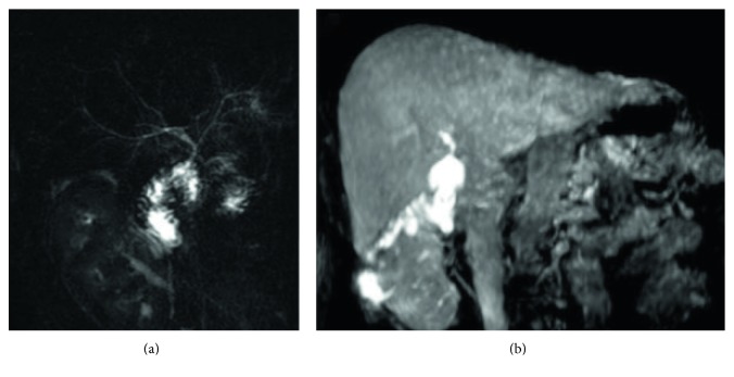
Figure 10.
Patient with a biliary-enteric anastomosis, performed after duodenopancreatectomy. 2D T2-weighted cholangiography (a) shows normal appearance of a biliojejunal anastomosis. Excretory phase—obtained after contrast administration of Gd-BOPTA—confirms regular opacification at the surgical connection (b).

