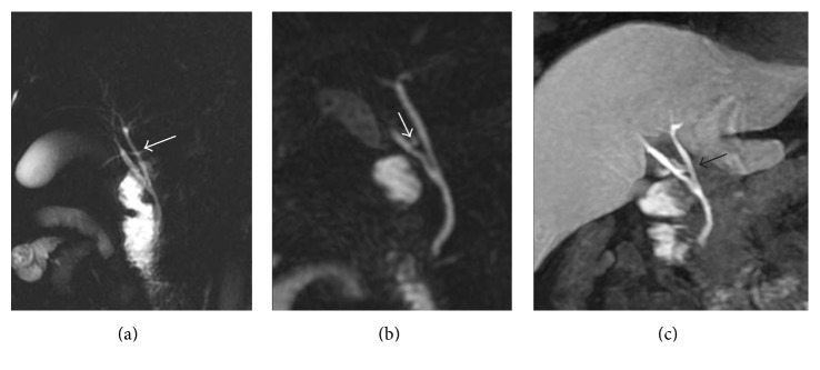
Figure 3.
Anatomical variations of the biliary tree. 2D thick-slab cholangiography and 3D MIP FRFSE ((a) and (b), resp.), show a caudal confluence between right and left biliary ducts; in addition, an aberrant right duct is suspected on (a) and (b) (white arrow). Gadoxetic acid-enhanced MRC clearly demonstrates the right aberrant duct and the cystic duct (black arrow), with a separate insertion along left biliary duct.

