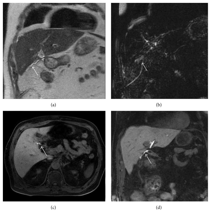
Figure 7.
Biliary leak after cholecystectomy. Coronal T2-weighted single-shot fast spin-echo image (a) and 3D FRFSE cholangiography (b) show a small fluid collection (white arrows in (a) and (b)). Images obtained in excretory phase ((c) and (d)) show progressive opacification (white arrows): a diagnosis of biliary leak was performed.

