Figure 5.

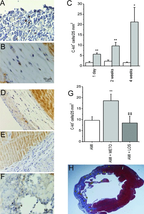
Metoprolol treatment for 2 weeks increases the number of c‐kit + cells in the border zone of the infarction. (A) c‐kit+ cells in rat sternum (positive control). (B) Single c‐kit+ cell in peri‐infarct area of the left ventricle. (C) Number of c‐kit+ cells in the anterior wall of LV of sham‐operated and AMI groups during the 4‐week follow‐up period. The area of counted section was determined by computerized methods and a number of positively staining cells was related to the area (cells/25 mm2). White bars indicate sham and gray bars AMI.*p < 0.05,**p < 0.01 vs. sham. Section of the left ventricle 2 weeks after AMI (D), AMI with metoprolol (E) and AMI with losartan (F). (G) Metoprolol treatment further increased the number of c‐kit+ cells in the LV.**p < 0.01 versus AMI,‡‡ p < 0.01 versus AMI + METO. (H) Section of LV 2 weeks after AMI with metoprolol treatment, yellow dots representing localization of c‐kit+ cells in the infarct border zone. Results are mean ± SEM (n= 5–6/group). AMI = acute myocardial infarction; METO = metoprolol; LOS = losartan.
