
Figure 7.
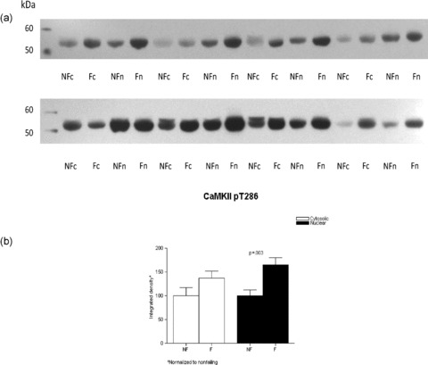
CaMKII phosphorylation is increased in the FHH. Lysates from eight nonfailing and eight failing LVs were loaded on a 10% SDS‐PAGE gel, the proteins fractionated and transferred to nitrocellulose membrane. The membranes were immunoblotted with a rabbit polyclonal antibody to CaMKII pT286, which has been shown to mirror total CaMKII activity. (A) The proteins were then visualized with enhanced chemiluminescence. Panel B illustrates the above results in graph form.