
Figure 2.
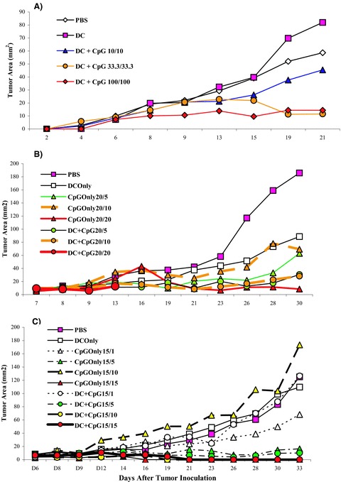
Dose‐finding studies of CpG DNA in combination with tumor lysate‐loaded DC in BALB/c mice bearing an established TS/A tumor. Individual groups of three BALB/c mice were inoculated subcutaneously in their right flanks with 5 × 10⁴ TS/A tumor cells. Tumors were allowed to grow for 7 days until they were palpable (approximately 1–3 mm2). (A) On day 7, post tumor implantation, TS/A tumor‐bearing mice were then treated twice, 7 days apart, by subcutaneous injections in the left flank with 100 μL of PBS, TS/A antigen‐loaded DCs alone (5 × 10⁵ DCs), or TS/A antigen‐loaded DC + CpG DNA at the test dose (nanomoles), where the DCs and CpG DNA were mixed in the same syringe just prior to injection. (B and C) CpG DNA alone was added as a control. Tumors were then measured with calipers twice a week in a blinded fashion, and tumor area (mm2) was determined by calculating the product of the two longest perpendicular diameters. Dosing of CpG DNA is represented as “x”/“y”, where “x” and “y” are the doses (nanomoles) of CpG DNA administered with the first and, 7 days later, the second injection, respectively. Note: the curves for 15/15 and 15/10 overlap.