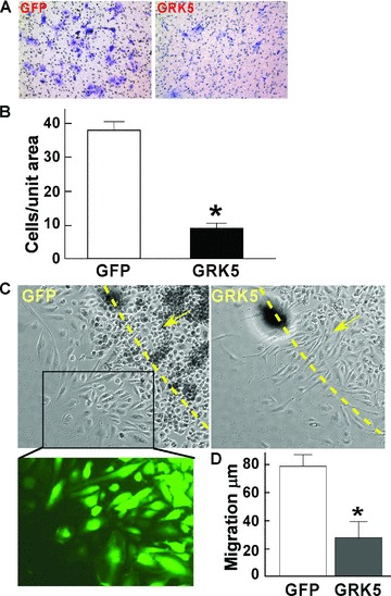
Figure 2.

GRK5 inhibits HCAEC migration. (A and B) HCAECs were infected with GRK5 or GFP adenovirus for 18 hours. Cells were then trypsinized and reseeded (1 · 104) in endothelial basal medium containing 0.5% FBS to the upper chamber of the Transwell system for 2 hours and then transferred to wells preloaded with 0.6 mL basal medium containing 0.5% FBS plus 20 ng/mL VEGF. Twenty‐four hours later, the inserts were fi xed and stained with crystal violet. The cells in the upper side of the insert were removed. Representative images of stained cells on the undersurface of the porous membrane as shown (A) were counted (n= 6) (B). (C and D) HCAECs were infected with GRK5 or GFP adenovirus as above and then trypsinized and reseeded in a cell clone cylinder for outward migration assay in the presence of VEGF (20 ng/mL). Representative images of cell migration and GFP expression are shown (C). Migration distances (μm) were measured 24 hours later (D) (n= 10). Data represent mean ± SD. *p < 0.001 versus control.
