Figure 1.

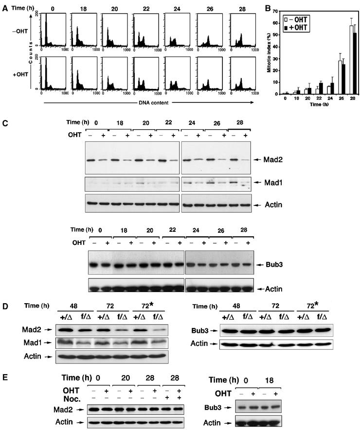
Loss of Trrap results in downregulation of Mad2 and Mad1 proteins. (A) Synchronization of Trrap-containing and Trrap-deficient cells. CER9 cells were synchronized at G0/G1 phase by serum starvation for 24 h with or without OHT pretreatment for 48 h. At indicated times after serum stimulation (i.e. 0 h corresponds to 48 h after addition of OHT), cells were stained with propidium iodide and analyzed by flow cytometry. (B) Cells were synchronized as in (A) and, after DAPI staining, the mitotic index was determined by scoring at least 100 cells. (C–E) Western blot analysis of mitotic checkpoint players in cells lacking Trrap. (C) CER9 cells were synchronized at G0/G1 phase by serum starvation for 24 h with or without OHT pretreatment for 48 h. Indicated times correspond to time after serum stimulation (i.e. 0 h corresponds to 48 h after addition of OHT). Protein extracts were prepared from samples taken at the indicated time points after serum stimulation and hybridized with anti-Mad2, anti-Mad1 and anti Bub3. Equal loading was verified by anti-actin antibody. (D) Primary MEFs of indicated genotypes were infected with adenovirus expressing Cre recombinase and at indicated time points after infection protein levels were determined by Western blotting. The asterisk (*) indicates that the cell cultures were incubated in the presence of nocodazole. (E) Western blot analysis of mitotic players in empty vector-transfected cells PSG1. Cell samples were taken at indicated time points after serum stimulation and protein levels of Mad2 and Bub3 were analyzed by Western blotting. Equal loading was verified by anti-actin antibody.
