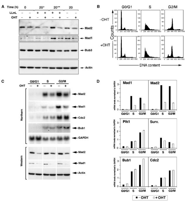
Figure 2.

Loss of Trrap compromises transcription of Mad2 and Mad1 genes and not their protein stability. (A) Reduced protein levels of Mad players in cells lacking Trrap are not due to increased protein degradation. CER9 cells were synchronized at G0/G1 phase by serum starvation for 24 h with or without OHT pretreatment for 48 h and released into the serum-containing medium in the presence or absence of the proteasome inhibitor LLnL (50 μM). An asterisk (*) and two asterisks (**) indicate that LLnL was added at the 0 and 16 h time point, respectively, after addition of serum. Cell lysates prepared from cells harvested at the indicated time points were analyzed by Western blotting. (B–D) Mad mRNA levels were downregulated in Trrap-deficient cells. (B) CER9 cells were grown in the presence or absence of OHT for 48 h and then synchronized at G0/G1 phase by serum starvation for 24 h. Synchronized S and G2/M populations were obtained by collecting cells at 18 and 28 h in the presence of nocodazole, respectively, after release from serum starvation, as evidenced by flow cytometry analysis. These populations were used for Northern blotting, Western blotting and ChIP assay (see below). (C) CER9 cells were synchronized as in (B), and total RNA and protein lysates were analyzed by Northern and Western blotting, respectively. RNA loading was controlled by GAPDH probe and protein loading was verified by anti-actin antibody. (D) Quantification of mRNA expression levels of the mitotic players. mRNA levels of indicated genes were analyzed densitometrically and normalized to GAPDH.
