
Figure 4.
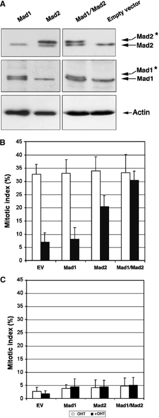
Ectopic expression of Mad1 and Mad2 in Trrap-deficient cells restores mitotic checkpoint. (A) Western blot analysis of protein expression of exogenous Mad1 and Mad2. CER9 cells were grown in the presence or absence of OHT for 24 h and then transfected by expression vector containing either Mad1 or Mad2 alone or Mad1 and Mad2 together. Note that exogenous human Mad1 and Mad2 proteins migrate at a slightly higher position on the gel (asterisk) in comparison to the corresponding endogenous mouse proteins. Actin was used as a loading control. (B, C) Ectopic expression of Mad proteins restores mitotic checkpoint. CER9 cells were grown in the presence or absence of OHT for 24 h and then cotransfected with an empty vector (EV) or indicated expression vectors and a vector expressing H2B-GFP. At 72 h after transfection, cells were incubated for further 10 h in the presence (B) or absence (C) of nocodazole. The mitotic index was determined by scoring GFP-positive cells with condensed chromosomes.