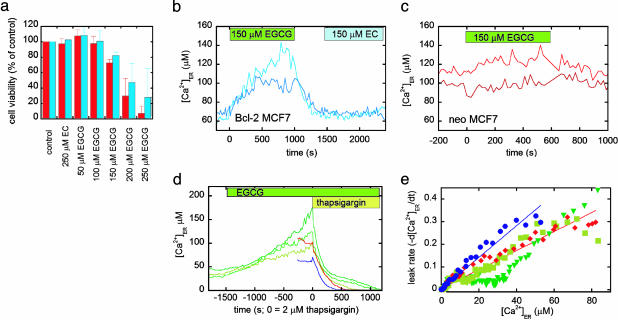
Fig. 5.
The effect of the green tea compound EGCG and a control compound EC on apoptosis and [Ca2+]ER of MCF-7 cells. (a) Cell viability (percent of the control), as determined by FACS analysis of propidium iodide and annexin V-stained neo (red) and Bcl-2 (blue) cells after 48-h treatment with nothing (control), EC, and increasing concentrations of EGCG. (b) Treatment of MCF-7 cells overexpressing Bcl-2 with EGCG and EC, as denoted by the green and blue boxes. The traces represent two different cells. (c) Treatment of MCF-7 neo cells with EGCG, as denoted by the green box. The traces represent two different cells. It should be noted that EC did not cause an increase in [Ca2+]ER in neo cells. (d) Bcl-2 cells treated with EGCG (green traces, each representing an individual cell) compared with neo (red) and Bcl-2 (blue) cells. At time t = 0, cells were treated with 2 μM thapsigargin in the absence of external Ca2+ to determine the ER Ca2+ leakage rate. (e) Comparison of the leakage rates of Ca2+ from the ER for Bcl-2 (blue circles), neo (red diamonds), and Bcl-2 cells pretreated with EGCG (green, upside-down triangles and squares), showing the decreased leakage rate upon EGCG treatment.

