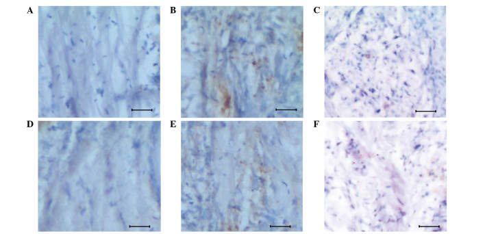
Figure 2.
Immunohistochemical variation observed between the normal and glioma tissues of mice. (A) Normal brain tissue of mice demonstrating a poor apoptotic signal. (B) Apoptotic signals in the glioma tissue. (C) Poor apoptotic signals in a DMOG-treated glioma mouse. (D) Normal brain tissue demonstrating reduced Egln3 expression. (E) Glioma tissue demonstrating increased expression of Egln3 protein. (F) Suppressed expression of Egln3 protein in the DMOG-treated glioma tissue. Scale, 100 µm. DMOG, dimethyloxalylglycine.

