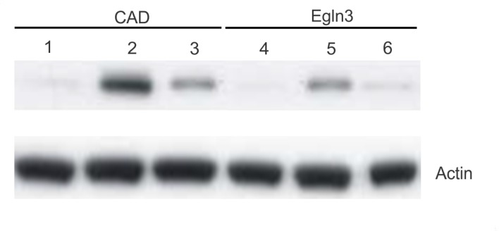
Figure 3.
Egln3 and apoptosis-specific CAD protein expression in the normal and glioma tissue. Lanes 1, 2 and 3 (CAD antibody) indicate the expression of apoptosis-specific CAD protein in the cell lysate of the normal, glioma and Egln3-suppressed glioma mouse tissue. Lanes 4, 5 and 6 indicate the expression of the Egln3 protein in the cell lysate of the normal, glioma and Egln3-suppressed glioma mouse tissue. The apoptotic signals were observed to be higher in the glioma tissue (lane 2) when compared with the normal brain tissue (lane 1) and Egln3-suppressed glioma mice tissue (lane 3). Similarly, Egln3 also exhibited a similar expression profile with increased expression in the glioma tissue when compared with the control and Egln3-suppressed glioma mice. The comparative analysis indicates the links between Egln3 and apoptotic protein expression. β-actin was used as a loading control. Egln3, Egl-9 family hypoxia-inducible factor 3; CAD, cell death-inducing DFF-like effector A.

