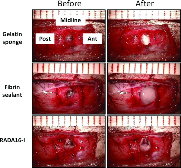
Figure 1.

Intraoperative photographs showing the cortical resection cavities before (left column) and after (right column) the applications of gelatin sponge, fibrin sealant and RADA16‐I. The cavities in the left column were temporarily filled with normal saline for clearer illustration. Ant, anterior; Post, posterior.
