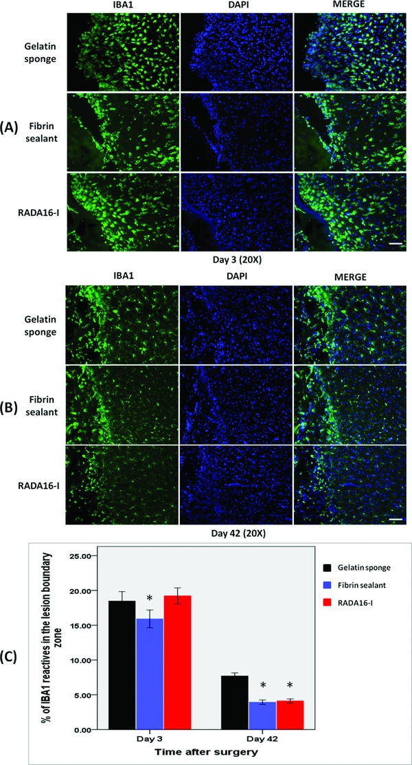
Figure 4.

Immunohistochemical studies showing microglial infiltration in the cerebral parenchyma adjacent to the lesion cavity on (A) day 3 and (B) day 42. (C) The degree of infiltration was more extensive after treatment with gelatin sponge than with fibrin sealant or RADA16‐I. *p < 0.001. Scale bar: A–B = 100 μm.
