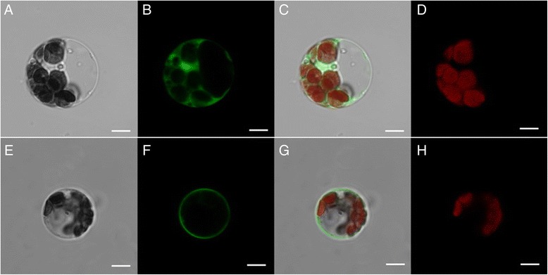
Fig. 5.

Subcellular localization of MeSTP7 in cassava mesophyll protoplasts. Transient expression of GFP, showing that the GFP is distributed throughout the nucleus and cytoplasm (a-d). The laser-scanning confocal microscopy images are the bright field image (a), fluorescence image (b), merged image (c) and autofluorescence image (d), respectively. The transient expression of GFP-fused MeSTP7 protein, showing that the MeSTP7-GFP fusion protein is likely localized to plasma membrane (e-h). The laser-scanning confocal microscopy images are the bright field image (e), fluorescence image (f), merged image (g) and chloroplast fluorescence (H), respectively. The bars = 5 μm.)
