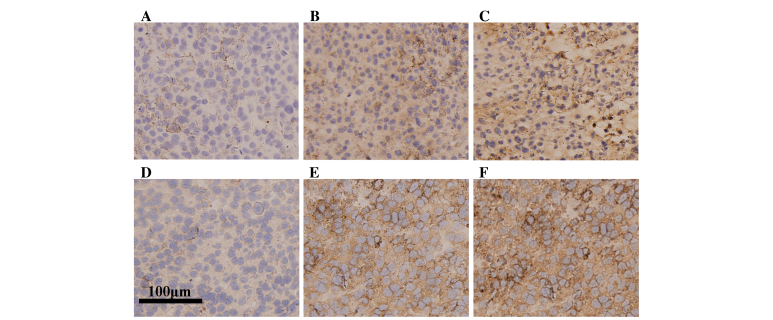
Figure 2.
Immunohistochemical staining of CD8a and CD4. CD8a immunostaining of tumors from (A) tumor group, (B) G-L group and (C) G-H group. CD4 staining of tumors from (D) tumor group, (E) G-L group and (F) G-H group. G-L group and G-H group exhibited a greater level of CD4+ or CD8a+ lymphocyte infiltration compared with the tumor group, and compared with the G-L group, the G-H group exhibited greater infiltration levels (magnification 1,000x; bar: 100 µm). G-L, low dose of ginsenoside Rh2; G-H, high dose of ginsenoside Rh2.

