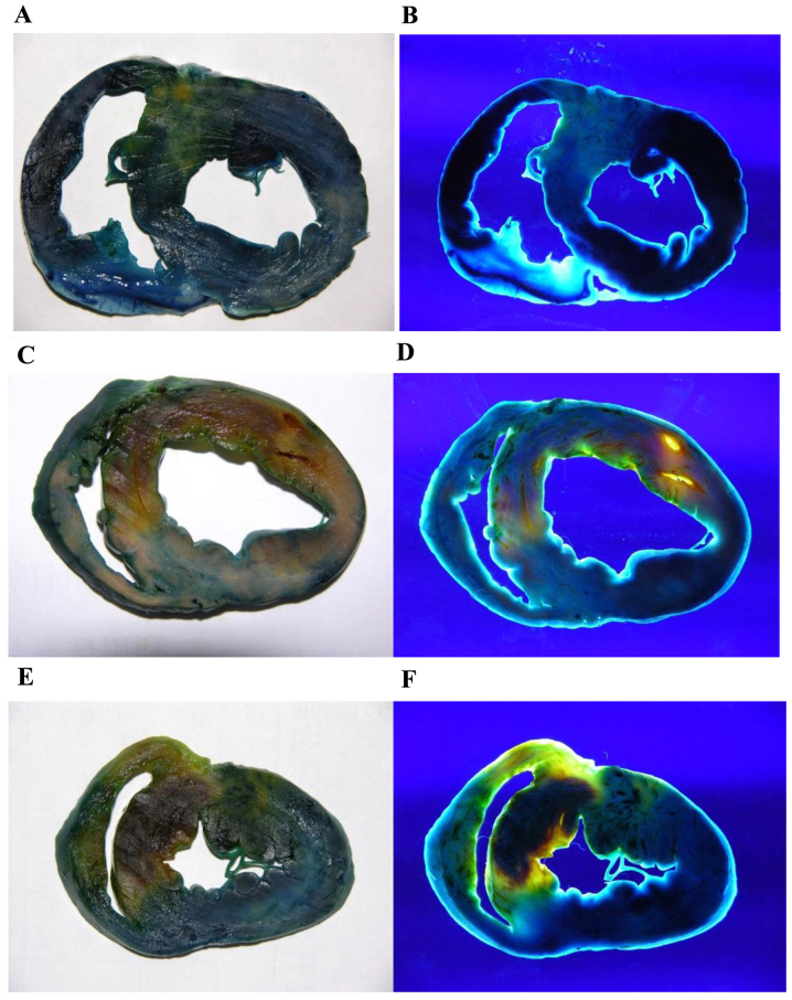
Figure 2.
Double staining of myocarium in the different groups (A and B) Sham-surgery (C and D) control and (E and F) PGE2 groups under natural (left column) and UV (right column) light. Normal myocardium, RA and NRA appeared dark blue, yellow, and dark red under natural light, respectively. Under UV light, the colors changed to black, bright yellow, and deep red, respectively. RAs of the PGE2 and control groups were not significantly different, whereas NRA of the PGE2 group was markedly smaller than of the control group. PGE2, prostaglandin E2; UV, ultraviolet; RA, reperfusion area; NRA, non-reflow area.

