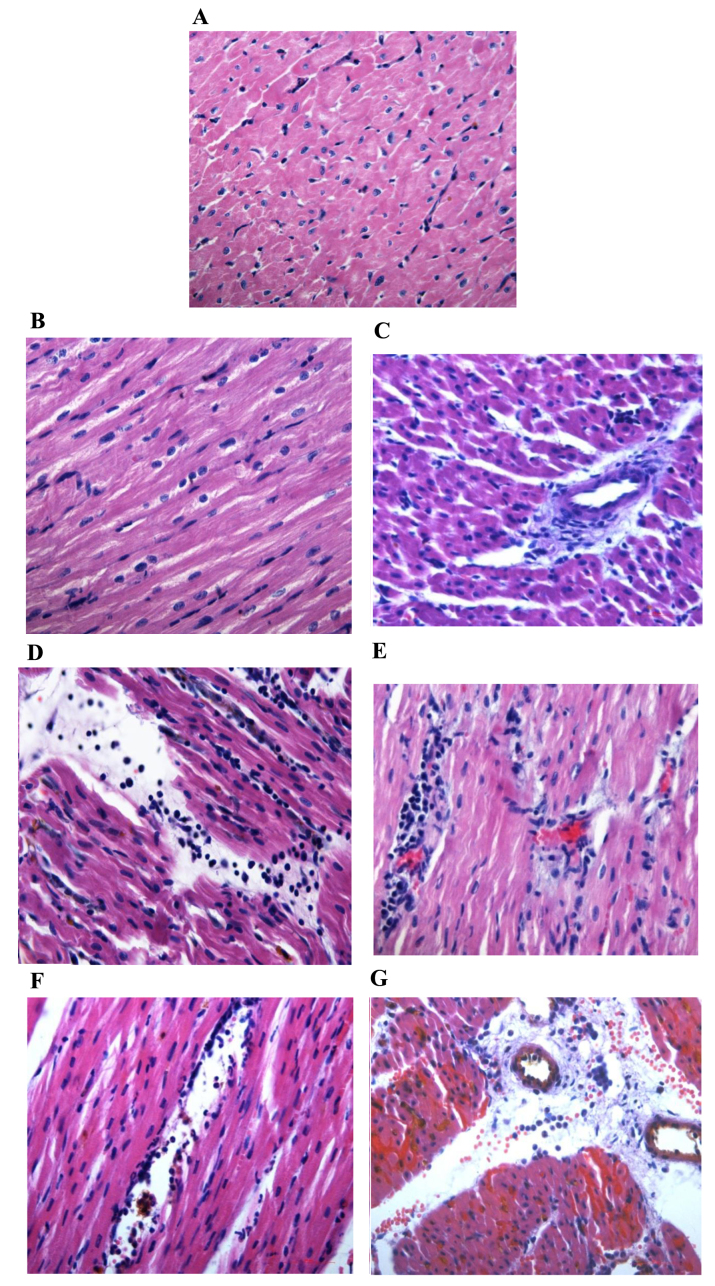
Figure 3.
Hematoxylin and eosin staining of myocarium in the different groups (magnification, ×400). Normal area of the three groups exhibited normal size cardiomyocytes, no hemorrhaging or neutrophil granulocyte infiltration. (A) Sham-surgery group, and normal areas of the (B) control and (C) PGE2 groups. The RA and NRA in the control group exhibited cardiomyocyte degeneration, hemorrhaging, edema, and significant interstitial neutrophil granulocyte infiltration compared with the normal area of the same group. (D) Reperfusion area and (E) NRA of the control group. No significant cardiomyocyte degeneration was identified in the RA and NRA in the PGE2 group, however, slight edema between the myocardial fibers, and mild neutrophil granulocyte infiltration was observed compared with the RA and NRA of the control group. (F) Reperfusion area and (G) NRA of the PGE2 group. The intravascular yellow staining is Thioflavin-S stain. PGE2, prostaglandin E2; RA, reperfusion area; NRA, no-reflow area.

