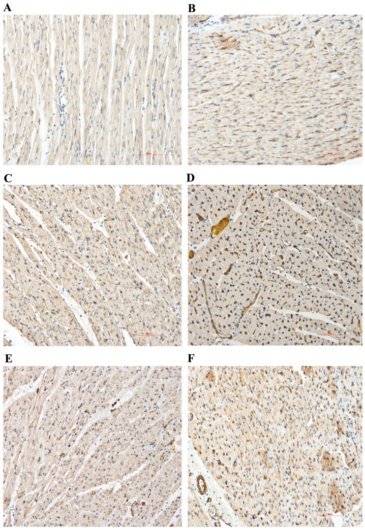
Figure 4.
Immunohistochemical analysis of the content and distribution of VEGF proteins (magnification, ×200). Myocardial VEGF protein expression levels of the control group markedly increased in the RA and NRA compared with the normal area. PGE2 markedly upregulated the expression level of VEGF in the RA and NRA further when compared with the control group. Normal area of the (A) control and (B) PGE2 groups; reperfusion area of the (C) control and (D) PGE2 groups; NRA of the (E) control and (F) PGE2 groups. VEGF, vascular endothelial growth factor; RA, reperfusion area; NRA, no-reflow area; PGE2, prostaglandin E2.

