
Figure 1. ATP triggers apoptosis of leukemia cells from AML patients via P2×7 activation.
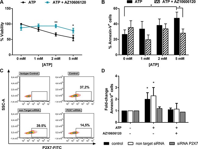
Leukemic cells isolated from AML patients were treated for 48 h with increasing doses of ATP, with or without (w/o) 10 μM AZ 10606120. Data are represented as mean +/− SEM (A) CellTiter 96 Aqueous One Solution assay was used to detect viability (n = 14) and (B) Annexin V/PI staining was used to detect apoptosis (n = 23). (C–D) To inhibit P2×7 expression, AML cells were nucleofected with a Non Targeting control siRNA or with P2×7-specific siRNA. After overnight, cells were treated with 5 mM ATP for 24 h, with or w/o 10 μM AZ 10606120 (n = 4). Results are expressed as fold-change of Annexin-V+ cells respect to untreated cells, for each group (% Annexin-V+ cells: 22.4 ± 7% control, 19 ± 6% Non Targeting Control siRNA, 23.4 ± 9.6% P2×7 siRNA). (C) Representative flow cytometric analysis of P2×7 expression after siRNA treatment. *p < 0.05.