
Figure 3. Stroma cells do not affect antineoplastic activity of ATP.
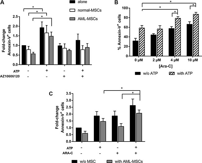
(A) AML cells were cultured alone or in presence of normal-MSCs and AML-MSCs stroma, at the ratio 10:1. After overnight, 5 mM ATP with or w/o 10 μM AZ 10606120 was added to the culture. Apoptosis induction was detected after 48 h by FACS analysis of Annexin-V+ cells. Results are expressed as fold change of Annexin-V+ cells, untreated AML cells cultured alone (39.6 ± 5.7 %) were used as reference and set as 1 (n = 7). (B) AML cells were treated for 48 h with increasing doses of ARA-C with or without 5 mM ATP and analyzed for apoptosis by flow cytometry (n = 6). (C) AML cells were cultured alone or in presence of AML-MSCs stroma, at the ratio 10:1. After overnight, 5 mM ATP with or w/o 4 μM ARA-C was added to the culture. Apoptosis induction was detected after 48 h. Results are expressed as fold change of Annexin-V+ cells, untreated AML cells cultured alone (Annexin-V+ cells: 31.6 ± 5.5%) were used as reference and set as 1. Data are represented as mean +/− SEM in all histograms (n = 6). *p < 0.05.