
Figure 5. HSCs were almost entirely unaffected by ATP treatment.
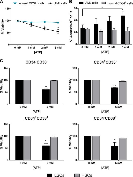
Leukemic cells isolated from AML patients and normal CD34+ isolated from healthy donors were treated for 48 h with increasing doses of ATP. (A) CellTiter 96 Aqueous One Solution assay was used to detect viability (AML n = 14, donor n = 4) and (B) Annexin V/PI staining was used to detect apoptosis (AML n = 23, donor n = 4). (C) CellTiter 96 Aqueous One Solution assay was used to detect viability of HSC and LSC subsets treated with 5 mM ATP for 48 h (AML n = 4, donor n = 3). Data are represented as mean +/− SEM in all histograms. *p < 0.05, **p < 0.01 significant with respect to untreated cells.