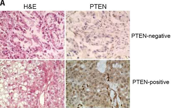
Figure 2. PTEN expression in primary OCCCs.

Using a validated antibody to PTEN, and previously reported scoring systems, PTEN expression was assessed in a panel of 50 primary OCCCs in a previously constructed tissue microarray. Cases were reviewed by at least two pathologists and discordant scores reassessed on whole tissue sections. Complete loss of PTEN expression was identified in 5/49 analysable cases (10%). Representative haematoxylin & eosin micrographs (left) and PTEN immunohistochemistry (right) of a PTEN-negative (top) and a PTEN-positive OCCC.
