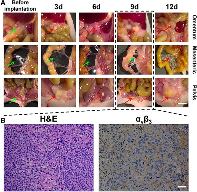
Figure 5. The dynamic and histological features of gastric cancer cell line SGC-7901 in peritoneal tumor formation.

(A) shows the morphological characteristics of tumors at different time points. The mesenteric tumor nodules at varying sizes were widely disseminated and can no longer be counted 12 days after implantation of tumor cells. Models at approximately the 9th day were selected, of which the green arrows indicate no pathological finding of tumor invasion ,while red arrows indicate cancerous tumor, Scale Bar = 5 mm; (B) shows the histological examination of three randomly selected tumor nodules from nude mice model at 9th day. It can be seen that there was a large number of abnormal cells and integrin αvβ3 was significantly expressed in the tumor tissue. Scale Bar = 50 μm.
