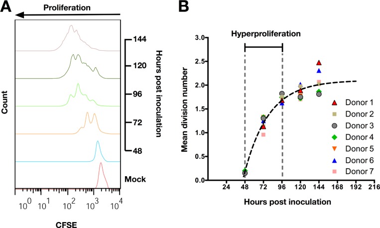
Figure 1. EBV induces hyperproliferation of tonsillar B-cells (TBCs) within 72 hours post inoculation.
(A) Representative data (Donor 7 from Figure 1B) of CFSE proliferation profile of TBCs at sequential days post EBV-B95.8 inoculation. Non-inoculated CD19+ B-cells harvested 120 hours post isolation (Mock) were used as negative control. (B) Mean division number of TBCs at sequential days post EBV-B95.8 inoculation. The function depicted as a black dashed line represents the average mean division number (MDN) for each time point measured. . Results shown are from TBCs from 7 donors.

