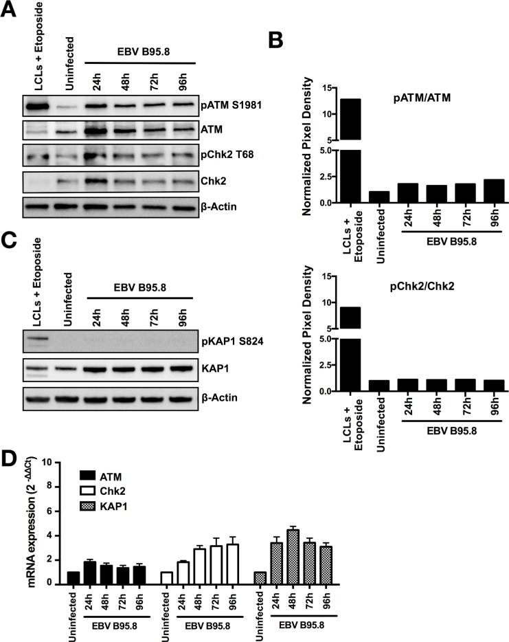
Figure 3. EBV inoculation does not elicit ATM/Chk2-mediated DDR activation in tonsillar B-cells (TBCs).
TBCs where inoculated with EBV-B95.8 (MOI 8). Non-inoculated TBCs at 24 hours post isolation were used as negative control (uninfected). Lymphoblastoid cell lines (LCLs) treated with Etoposide for 6 hours served as positive control. (A) Cells were harvested at the indicated time points, and expression of total ATM or Chk2 and pATM S1981 or pChk2 T68 was analyzed by western blotting. (B) ATM and Chk2 phosphorylation and total protein amount were quantified by densitometric analysis. Data are represented as ratio of phosphorylated-to-total. Quantification was performed using the software imageJ 1.49t. (C) Total KAP1 and pKAP1 S824 was analyzed by western blotting. The results shown are representative of 3 independent experiments. (D) ATM, Chk2 and KAP1 mRNA expression was determined in EBV-inoculated B-cells at the indicated time points by RTqPCR relative to 18 S RNA and shown as fold change relative to non-inoculated cells. Data are presented as mean ± SEM of 4 donors.

