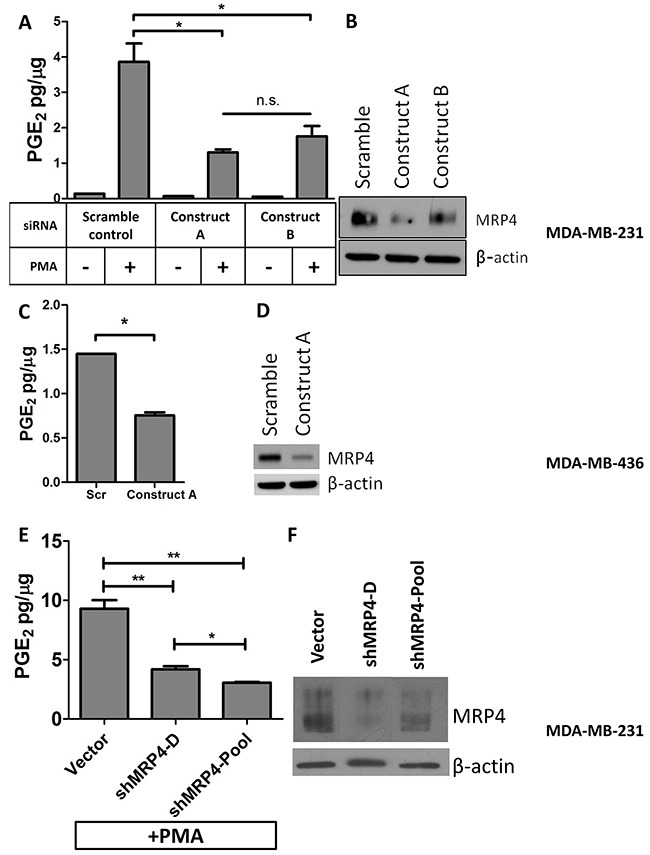
Figure 2. Knockdown of MRP4 in MDA-MB-231 and MDA-MB-436 cells suppresses the export of PGE2.

MDA-MB-231 cells were transfected with MRP4 siRNA (3 nmol/L) for 24 hours before being stimulated with PMA (80 nmol/L, 1 hr) and replacing growth media. After 16 hours, conditioned media and total protein lysate was collected. A. Conditioned media from MRP4-silenced cells (MDA-MB-231) stimulated with PMA was assayed for PGE2 and total protein. B. A representative western blot of MDA-MB-231 cells transfected with siRNA (3 nmol/L) and stimulated with PMA shows decreased expression of MRP4. MDA-MB-436 cells were transfected with MRP4 siRNA (10 nmol/L). C. Conditioned media from MDA-MB-436 cells was collected after overnight incubation and assayed for PGE2 content. D. A representative western blot showing 68% decreased MRP4 expression following siRNA transfection of MDA-MB-436 cells. E. MDA-MB-231 vector control, clone (shMRP4-D), and pool (shMRP4-Pool) populations of stable MRP4 knockdown cells were briefly stimulated with 80 nM PMA and incubated overnight in fresh growth medium. Conditioned media was collected and assayed for PGE2. F. A representative western blot showing decreased MRP4 expression of MDA-MB-231 clone shMRP4-D and shMRP4-Pool cell lines compared to vector control cells. PGE2 is expressed as mean ± SEM pg/μg protein from triplicate determinations. β-actin was used as a loading control.* p < 0.05, ** p < 0.01, n.s. = not significant.
