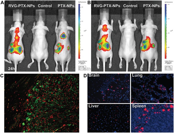
Figure 7. In vivo images were taken at 24.

A. and 48 B. hours in live animals after administration of DiSC3(5) labelled RVG-PTX-NPs to mice via tail vein. Significant amount of fluorescence signals appeared in the head after 24 and 48 hours. The brain sections from human glioma of SCID mouse model were labeled with IBa-1 and GFP to identify TAMs/microglia (C, red) and human glioma U87 cells (C, green). The frozen sections was mounted with DAPI (blue) to examine the tissue distribution of DiSC3(5) labelled RVG-PTX-NPs (D, red).
