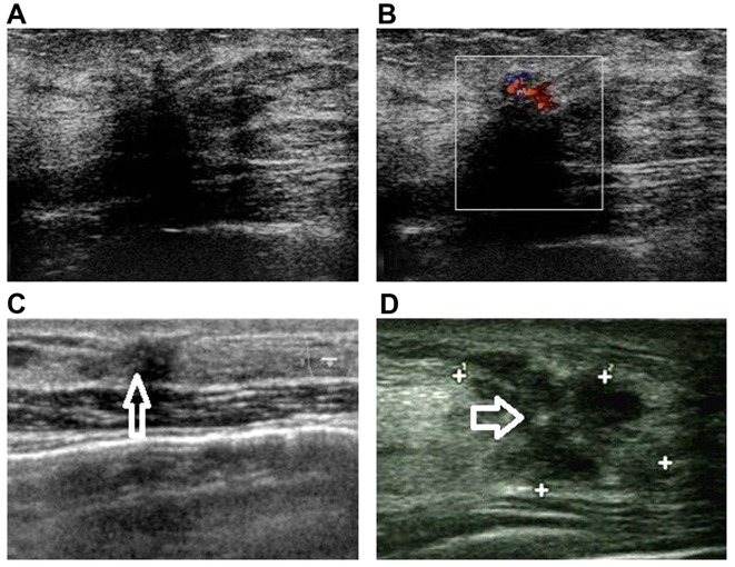
Figure 2.
Malignant signs on imaging. (A) Hypoechoic mass with an irregular shape, indistinct and angled margin [Breast Imaging Reporting and Data System (BI-RADS) category 4C], which was confirmed as complex sclerosing adenosis (SA). (B) Heterogeneously echogenic area with rich blood supply (BI-RADS 4B), confirmed as SA with ductal epithelial hyperplasia. (C) Burr-like mass with diffusely punctate calcifications (arrow) (BI-RADS 4B), diagnosed as SA with calcium deposition. (D) Irregular mass with angled margin and clustered amorphous calcifications (arrow) (BI-RADS 4B), confirmed as SA with atypical ductal hyperplasia and calcium deposition.

