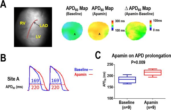
Figure 1.
Effect of apamin on APD80 prolongation in HF ventricles. A, APD80 map before and after apamin infusion in a failing heart. The ΔAPD80 map shows a heterogeneous distribution of APD prolongation with apamin. B, APD80 trace before (blue line) and after (red line) apamin infusion at pacing cycle length (PCL) 300ms. C, Summary data shows that the APD80 and was significantly prolonged after apamin infusion in HF ventricles.

