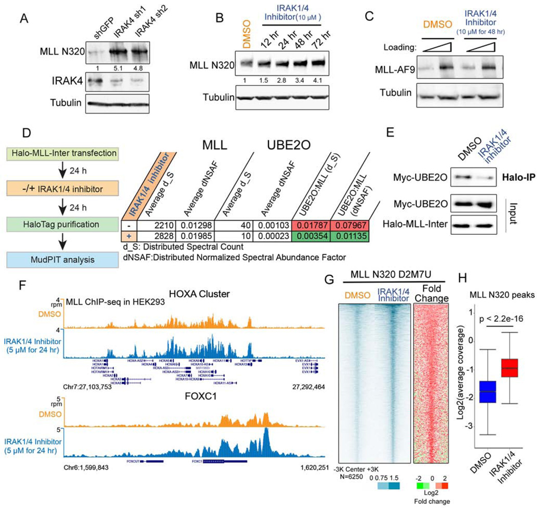
Figure 3. IRAK inhibition stabilizes MLL protein and increases genome-wide MLL occupancy.
(A) IRAK4 knockdown leads to increased levels of endogenous MLL protein in HEK293 cells. Fold changes of MLL N320 protein relative to shGFP are indicated.
(B) IRAK1/4 inhibitor stabilizes endogenous MLL protein. HEK293 cells were treated with 10 µM IRAK1/4 inhibitor for the indicated times. Fold changes of MLL N320 protein relative to vehicle (DMSO) are indicated.
(C) IRAK1/4 inhibitor has no obvious effect on MLL-AF9 stabilization. Flag-MLL-AF9 HEK293 cells were treated with 10 µM IRAK1/4 inhibitor for 2 days and subjected to western blotting with the FLAG monoclonal antibody.
(D–E) IRAK1/4 inhibitor decreases MLL-UBE2O interaction. Halo-MLL-Inter transfected HEK293 cells were treated with 10 µM IRAK1/4 inhibitor 24 h, purified with HaloLink resin and subjected to MudPIT analysis.
(F) Genome browser tracks of MLL-N320 D2M7U ChIP-seq at HOXA and FOXC1 loci after DMSO or IRAK1/4 inhibitor treatment for 24 h. IRAK1/4 inhibitor increases MLL occupancy at HOXA and HOXC loci.
(G) Heat map analysis of MLL occupancy after IRAK1/4 inhibitor treatment in HEK293 cells. Each row represents a peak of MLL occupancy (N=6250), with rows ordered by decreasing MLL occupancy in the inhibitor-treated condition.
(H) MLL occupancy is significantly increased after IRAK1/4 inhibitor treatment. Box plots depict the read coverage for all MLL peaks. The p value was calculated with the Wilcoxon signed-rank test.
See also Figures S3 and Table S1.

