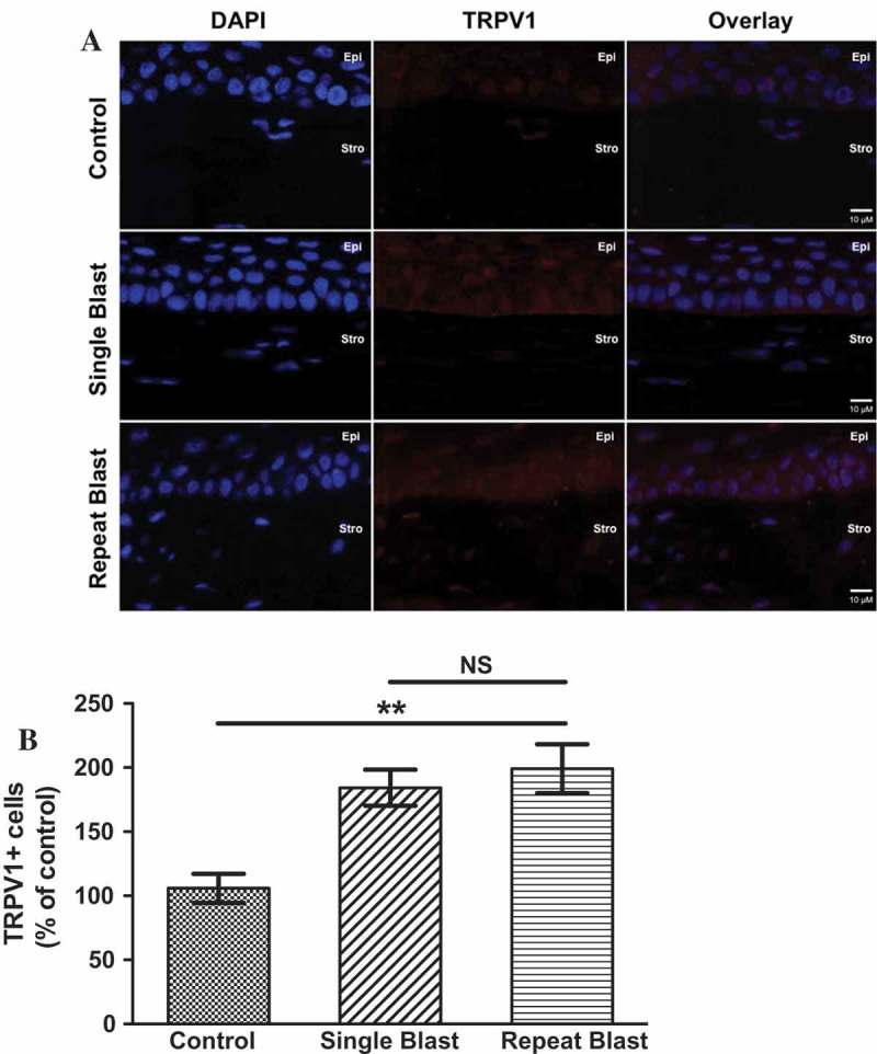
Figure 2.

(A) Immunofluorescence analysis was performed on rat corneal sections exposed to either a single or repeated blast (70 KPa). Tissues were subjected to staining with an anti-TRPV1 (1:250) antibody and probed with an Alexa Fluor 568 secondary antibody. Nuclei were visualized with DAPI staining (1:1000). Images were captured at 60× magnification, oil immersion. (B) Quantification of TRPV1-positive cells as a percent of control animals. One-way ANOVA, Bonferroni post-hoc analysis, **p < 0.01, NS not significant. Cornea layers are indicated as Epi = epithelial, Str = Stromal.
