
Figure 4.
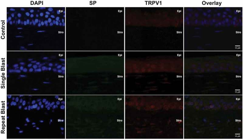
Immunofluorescence analysis was performed on rat corneal sections exposed to either a single or repeated blast (70 KPa). Tissues were subjected to staining with anti-TRPV1 (1:250) and anti-SP (1:500) antibodies and probed with Alexa Fluor 568 and 488 secondary antibodies, respectively. Nuclei were visualized with DAPI staining (1:1000). Images were captured at 60× magnification, oil immersion. Cornea layers are indicated as Epi = epithelial, Str = Stromal.