Figure 1.
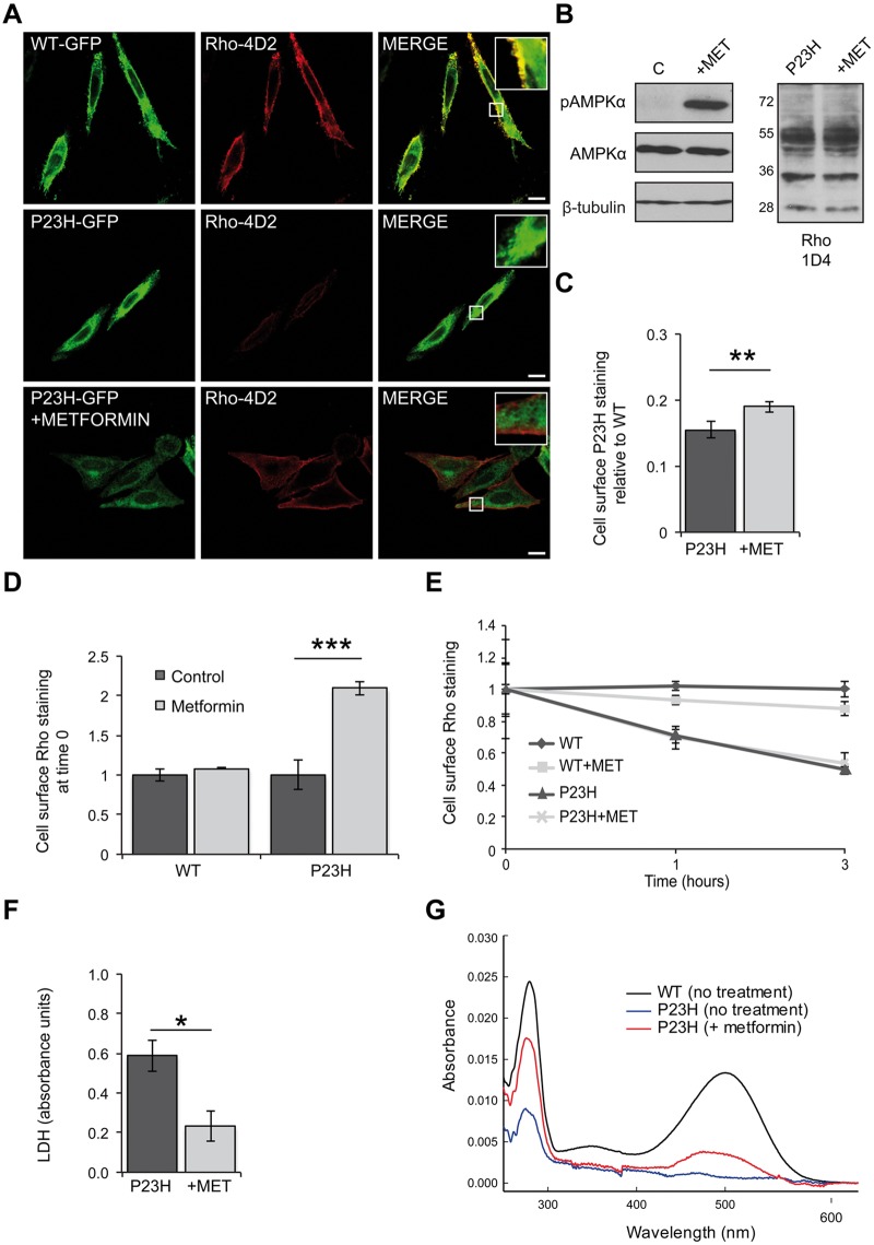
Metformin improves P23H rod opsin traffic and folding and reduces P23H-induced cell death. (A) SK-N-SH cells transfected with WT-GFP rod opsin (green) or P23H-GFP rod opsin (green) were treated with metformin (1 mM) for 18 h. Fixed, non-permeabilised cells were stained with Rho-4D2 antibody against the extracellular N-terminus (red). Confocal microscopy imaging under identical conditions. Scale bar: 10 μm. Boxed regions show higher magnification. (B) Immunoblot with an anti-phospho-AMPKα (p-AMPKα) or an anti-AMPKα antibody. β-Tubulin was used as a loading control. Untreated- (C) and metformin- (+MET) treated P23H-GFP cells were blotted with the Rho-1D4 antibody. (C) In-cell western analysis of HA-P23H rod opsin. SK-N-SH cells were fixed and immunostained with an HA antibody against the extracellular N-terminus of rod opsin. The non-permeabilised (cell surface) immunoreactivity was determined as a percentage of total permeabilised immunoreactivity. The data were normalised to the amount of cell surface HA-WT rod opsin, values ± SEM, n ≥ 4, **P < 0.01, unpaired two-sided Student's t test. (D-E) Rhodopsin internalisation assay in HA-WT or HA-P23H SK-N-SH untreated cells or metformin treated (18 h). Live cells were incubated with an HA antibody for 30 min at 37 °C, washed, media returned and rod opsin was allowed to internalise for 0, 1 and 3 h. (D) Cell surface rod opsin staining at time 0. Values ± SEM, n ≥ 3, ***P < 0.001, unpaired two-sided Student's t test. (E) Remaining cell surface WT or P23H rod opsin staining after 0, 1 and 3 h of internalisation in the presence or absence of metformin, normalised to time 0. Values ± 2SEM, n ≥ 3. (F) LDH assay on cells expressing P23H-GFP rod opsin treated with 1 mM metformin (+MET). Average absorbance values ± SEM, n ≥ 3, *P < 0.05, unpaired two-sided Student's t test. The LDH positive control (100% cell death) was 2.3 absorbance units. (G) UV-visible absorption spectra of immunoaffinity purified WT (black) and P23H (blue) rhodopsin pigments obtained from tetracycline inducible HEK293S cell lines treated with 300 μM metformin (red) for 24 h during expression The WT sample was diluted to show the profile on a similar scale.

