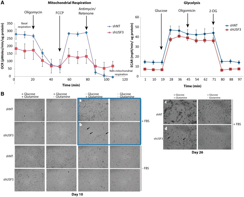
Figure 5.
USF3 knock down cells had impaired mitochondrial respiration and altered glutamine usage. (A) Seahorse mitochondrial metabolic profiling. Both oxygen consumption rate (OCR, lest panel) and extracellular acidification rate (ECAR, right panel) were measured in shNT and shUSF3 NThy-ori 3-1 cells. (B) Phase-contrast microscopy images of shNT or shUSF3 NThy-ori 3-1 cells cultured in different combination of culture medium for 10 days (left panel), or extended to 26 days with FBS (right panel). As highlighted in the blue square, when cultured without glucose but with addition of glutamine, shNT cells showed mainly dotted morphology (panel a) indicating dying cells, while more shUSF3 cells (panel b) exhibited normal attached cell shape (pointed by arrows) suggesting survival. Similar observations lasted even after 26 days under the same culture conditions (panel c and d) where there were a lot more live shUSF3 cells in panel d starting to contact each other and form cellular matrix.

