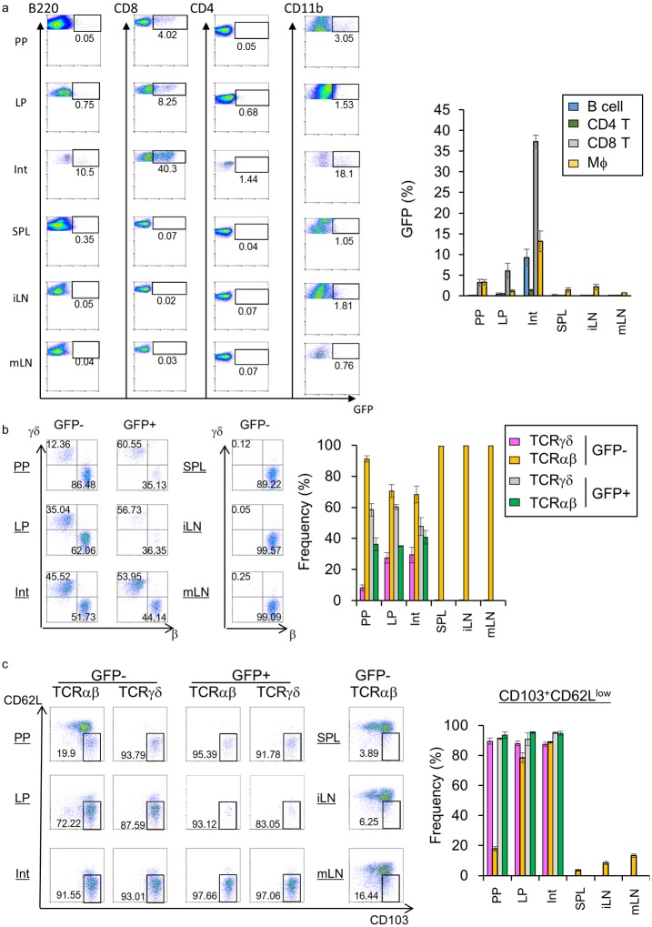
Fig 1. Expression of Opn in specific CD8+ T cell subsets in the intestine, but not in other secondary lymphoid organs.
Six-week-old female EGFP-Opn mice were analyzed by flow cytometry. Cells obtained from Peyer’s patches (PP), lamina propria (LP), intestinal epithelium (Int), spleen (SPL), inguinal (iLN), and mesenteric lymph nodes (mLN) were stained for detection of (a) CD4 (CD3+CD4+), CD8 T cells (CD3+CD8α+), B cells (B220+), and macrophages (CD11b+) (b) TCRαβ or TCRγδ+ cells in CD3+CD8α+ cells (c) CD103+CD62Llow resident memory-type CD8 T cells. Graphs show (a) GFP+ cells, (b) TCRαβ+ and TCRγδ+, and (c) CD103+CD62Llow TCRαβ+ or TCRγδ+ cells in GFP+ or GFP- populations. Bars indicate ±S.E.M (n = 3~5). Data are representative of two independent experiments.

