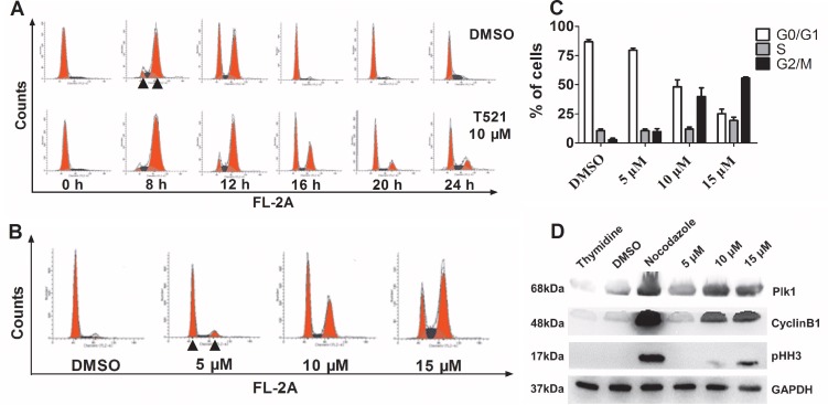
Figure 3. T521 induces mitotic arrest in HeLa cells.
A. HeLa cells (1.0×10⁵ cell/well) were synchronized at the G1/S boundary by double-thymidine block, and then released into medium with or without T521 (10 μM). Cell cycle progression was analyzed for DNA content by flow cytometry. DNA contents of 2C (left) and 4C (right) are indicated by triangles in the image. B. The G2/M arrest induced by different concentrations of T521 after release from double-thymidine block for 16 hrs. Cell cycle analysis was performed as described in A. C. Quantitative analysis of cell cycle distribution of DMSO or T521-treated HeLa cells in panel B (n=3). Error bars represent SD. D. Protein levels of mitotic markers in T521-treated HeLa cells. Synchronized HeLa cells were released into medium containing T521 at the indicated concentrations or nocodazole (50 ng/mL) for additional 16 hrs. Cellular extracts were prepared and the protein levels of Plk1, Cyclin B1 and pHH-3 were analyzed by western blotting. Nocodazole-treated cells were used as a control for mitotic arrest. DMSO-treated cells and cells synchronized with thymidine (2 mM) were also used as controls. GAPDH was used as the loading control.

