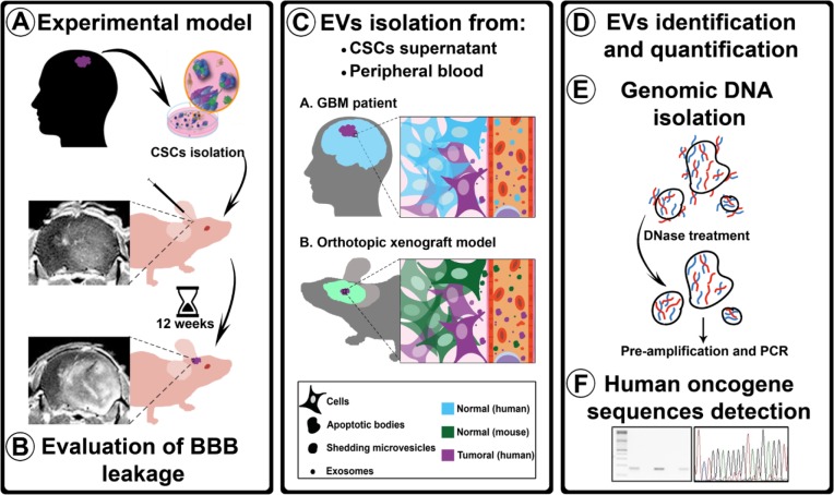
Figure 1. Experimental procedure flowchart.
A. Isolated hCSCs from 2 GBM patients were xenotransplanted in athymic mice. After 12 weeks, the animals were transcardially perfused. B. BBB permeability was evaluated using three assays: MRI, Evans Blue staining, and albumin extravasation. C. EVs (ABs, SMVs, and EXOs) were isolated from hCSCs-enriched culture supernatants and from mouse peripheral blood. D. EVs were identified using TEM, tracking analysis, and CD63 tetraspanin quantification. E. To ensure that the analyzed DNA was confined within the EVs, supernatants and plasma were treated with DNase before gDNA isolation; after the isolation, gDNA was pre-amplified before performing PCR analysis with human-specific primers. F. Sequences detected were sequenced to confirm their human origin.

