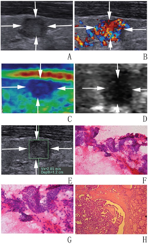
Figure 2. Images in a 32-year-old woman with papillary mirocarcinoma.

A, B. at conventional US, an 8-mm thyroid nodule (arrows) in the right lobe of the thyroid appears to have moderate hypoechogenicity, poorly defined margin, irregular shape, and rich blood flow. At elastography, C. CSE score of 4 (arrows), D. ARFI imaging grade of VI (arrows), and E. SWS of “2.85” m/s are assigned. F, G. FNA cytology (haematoxylin-eosin stain, original magnification, ×200 (F), ×400 (G)) shows part follicle cells with nuclear atypia and the nodule is diagnosed as AUS/FLUS. H. Histologic specimen (hematoxylin-eosin stain; original magnification, ×50) shows that this thyroid nodule is finally confirmed to be a papillary mirocarcinoma.
