
Figure 3. Images in a 61-year-old woman with Hashimoto nodule.
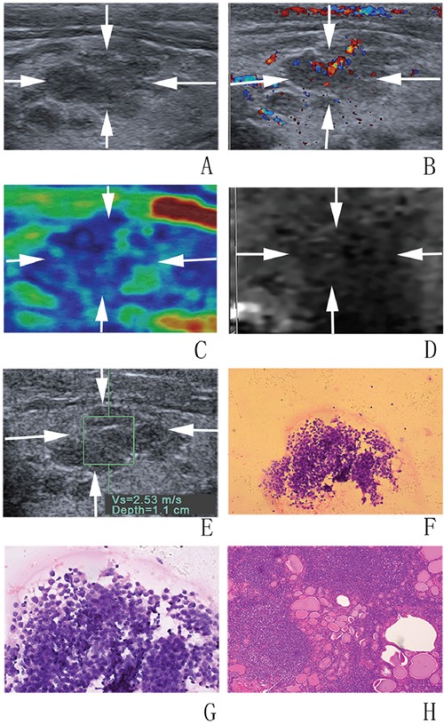
A, B. at conventional US, a 12-mm thyroid nodule (arrows) in the right lobe of the thyroid appears to have coarse echogenicity background, moderate hypoechogenicity, poorly defined margin, irregular shape, and rich blood flow. At elastography, C. CSE score of 3 (arrows), D. ARFI imaging grade of III (arrows), and E. SWS of “2.53” m/s are assigned. F, G. FNA cytology (haematoxylin-eosin stain, original magnification, ×200 (F), ×400 (G)) shows a few follicle cells with architectural atypia under the background of lymphocytes and diagnosed as AUS/FLUS. H. Histologic specimen (hematoxylin-eosin stain; original magnification, ×100) shows that this thyroid nodule is finally confirmed to be a Hashimoto nodule.