Figure 6.

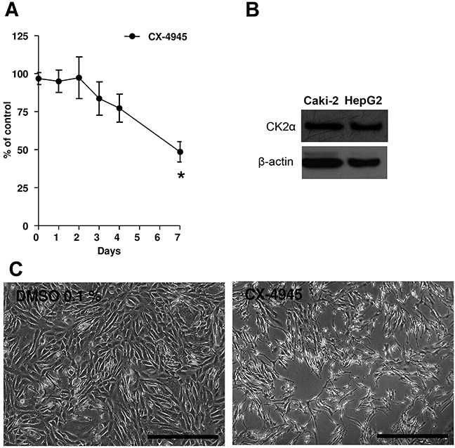
A. Caki-2 cells were treated with CX-4945 (10 μM) for 7 days. Experiments were repeated three times and data (absorption, ABS) were expressed as the means ± SEM of 3 replicates for each condition. Absorbance values were normalized to vehicle (DMSO). Student's T-test was used for statistical comparison of data sets at any given time point. *p< 0.01 vs. Control (vehicle). B. Western blot analyses of CK2α in Caki-2 cell lysates and HepG2 not treated with CX-4945. HepG2 served as a positive control. Actin expression served as a loading control. C. Pictures showing Caki-2 cells at the seventh day of the proliferation assay. At day 7, vehicle (DMSO 0.1%) was confluent, while cells in the presence of CX-4945 (10 μM, right picture) show a reduction to 49%. The scale bar in each picture corresponds to 500 μm.
