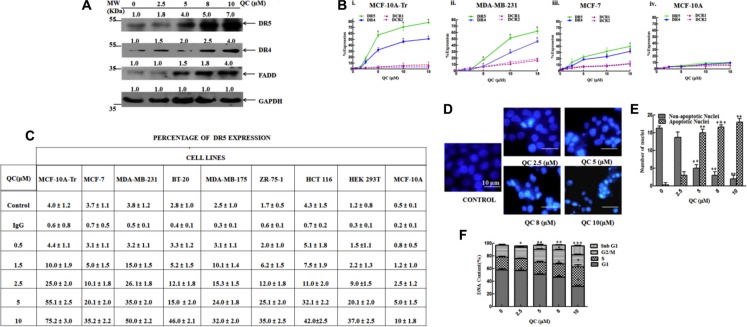
Figure 2. QC caused apoptosis in cancer cells by increasing the expressions of death receptors.
(A) Expressions of proteins in QC treated MCF-10A-Tr cells. (B) Expressions of different receptors in cell surface measured by FACS. i) MCF-10A-Tr, ii) MDA-MB-231, iii) MCF-7, and iv) MCF-10A cells. (C) Expression of DR5 in different cells after QC exposure. (D) Apoptosis measured by DAPI staining. (E) Graph representing the number of apoptotic and non-apoptotic nuclei in Figure 2D. (F) Measurement of apoptosis by FACS analysis of MCF-10A-Tr cells treated with QC. The images shown are representative of three different experiments. Data are the mean ± SD of three different experiments.

