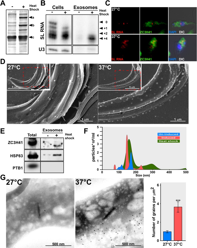
Fig 8. SL RNA accumulates during heat-shock and is secreted by exosomes.
(A) Induction of heat-shock proteins. Cells (107), were incubated at either 27°C or 37°C (heat-shock) for 40 minutes in Methionine-free medium; 35S-Methionine (100 μCi) was added for 10 min followed by a 5 min chase. The cells were collected, and the proteins were separated on a 10%-SDS gel and subjected to autoradiography.a-HSP83 b-HSP70 c-tubulin. (B) SL RNA accumulates under heat-shock. Cells (108 in 10 ml) were incubated at 27°C or 37°C, for 1h. RNA was prepared from semi-purified exosomes and analyzed by primer extension. The SL RNA cap-4 modifications are indicated. (C) SL RNA and ZC3H41 are transported from the nucleus to the cytoplasm under heat-shock. Cells were incubated for 1hr at the temperature indicated and subjected to in situ hybridization. Nuclei were stained with DAPI. The merge was performed between IFA, in situ hybridization and DAPI staining. (D) SEM analysis of un-induced cells, and cells exposed to heat-shock. Cells were either incubated at 27°C or 37°C (heat-shock) for 1 hour. After incubation, the cells were fixed and visualized under EM; the scale bars and the treatment of the cells are indicated. (E) Exosomes are secreted under heat-shock. Cells (108) were incubated at either 27°C or 37°C (heat-shock) for 40 min, and exosomes were prepared as described in Materials and Methods, and subjected to western analysis with the indicated antibodies. (F) Quantitation of the exosomes secreted under heat-shock. Exosomes from wild-type, SmD1 silenced cells and heat-shocked cells were analyzed by NanoSight. Exosomes from un-induced cells (blue), SmD1 silenced cells (red), and heat-shock (green). (G) SEM-Immunogold to detect ZC3H41 secretion under heat-shock. Wild-type cells were incubated for 1h at the indicated temperature. Cells were subjected to immunogold staining. The backscatter images are presented; scale bars are indicated. The statistical analysis represents the mean ± S.E.M **P <0.01, and ***P <0.005 compared to–Tet, using Student's t-test.

