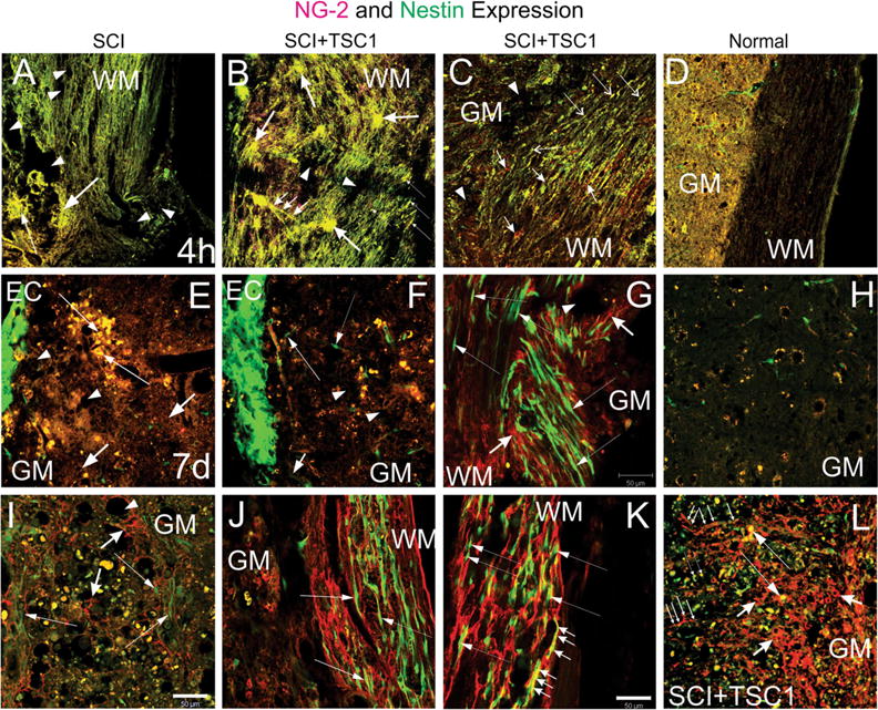
Fig. 4.

Two types of progenitors were present upon TSC1 intervention. a and b show low magnification views of the lesion epicenter. a Four hours after injury at the level of the lesion, the tissue appeared disrupted and without defined cellular structures that would have expressed nestin (neural progenitors) or OLPs identified by NG2. Moreover, the tissue showed a spongy appearance (arrows), and presented extensive cavitation (arrowheads). b In contrast the tissue from mice receiving TSC1 showed two types of structures: 1 Clusters of cells co-expressing nestin and NG2 (yellow). These cells were characterized by extending several long cell processes that appeared to go above nestin negative NG2 positive structures as a bridge for small cells to migrate towards the WM (small arrows). 2 Cells devoid of their processes (thin, long arrows). There were very small signs of cavitation (dotted areas). c Below the lesion (1.5 mm) along white matter tracks numerous small bipolar nestinansd NG2 expressing cells had appeared (Long and thin arrows). There were also interfascicular cells expressing solely NG2 mainly in their cell body (short arrows). d Low magnification view of a spinal cord of a naïve mouse at the T12 level. Seven days after injury panels e–g. e At higher magnification, the ependymal canal showed intense nestin expression (EC). The tissue adjacent to the ependymal canal had a spongy appearance (short arrows). A few patches of cells co-expressing nestin and NG2 were visible (yellow cells; long, thin arrows). At a longer distance from the EC the yellow cells were disappearing leaving behind only debris. f Seven days after injury and TSC1 treatment, the ependymal canal displayed an intense nestin expression. Beyond the EC few scattered, bipolar nestin-bearing cells appeared to migrate perpendicular to the EC and towards the white matter (long thin arrow). There was some cavitation in the GM (arrowheads). Some NG2 and nestin co-expressing cells presumably had migrated away from the canal (yellow cells). g The WM displayed a plethora of nestin-expressing bipolar cells mostly arranged in rows following the direction of the axons. Thus, nestin-expressing cells that had migrated following those axons directly exposed to the severe crush accumulated where the axons were severed. Long, thin arrows show examples of bipolar cells appearing to migrate along the axons. In areas where the white matter suffered to a lesser extent, nestin-expressing cells with elongated thin processes were also seen (thin, long arrows). Cavitation was less frequent in TSC1 treated mice (arrowhead). NG2 expressing cells had formed a matrix with their cell processes that intermingled with the axons and nestin-expressing cells (short, thick arrows). h Low magnification view of a naïve spinal cord’s gray matter. i Fourteen days after the intervention, the GM of mice that did not receive TSC1 had a lace-like aspect and NG2 labeled fibrous cell processes surrounding the cavities that formed upon tissue loss. Numerous cells that co-expressed both NG2 and nestin were enlarged and seemed to be dying. Very few green cells bearing faint nestin expression were visible at this time point (long, thin arrows). In contrast, mice treated with TSC1 (panel L) was populated of numerous NG2-expressing cells with large cell body and fibrous processes (short, thick arrows) Long. They had formed a dense structure across the GM that appeared to serve as a migratory substrate of both nestin expressing cells (long thin arrows) and NG2/nestin co-expressing cells (thin, long arrows). NG2 expressing cells were much larger than the NG2/nestin co-expressing cells. j Surprisingly, the white matter of mice 14 days after SCI had preserved its structure to some extent and nestin positive cells had reached them and appeared to migrate along axons (thin, long arrows). k In mice treated with TSC1 the white matter was populated by bipolar nestin positive cells intermingled with NG2 intensely labeled cells, the WM structure was nicely preserved allowing for the migration of nestin-expressing neural progenitors (thin, long arrows). Interestingly, bipolar cells co-expressing nestin and NG2 were abundant (small arrows), while in mice without TSC1 bipolar cells in the white matter mostly expressed nestin alone
