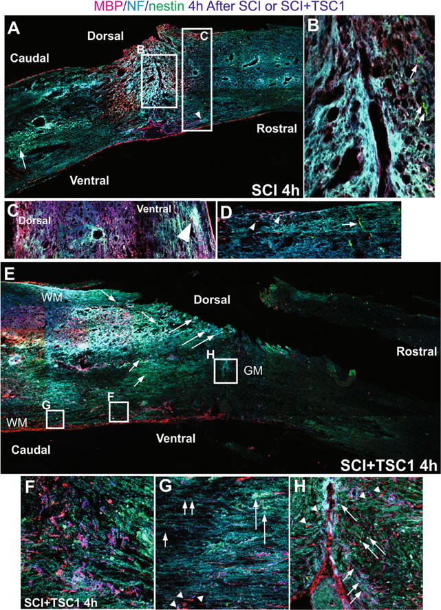
Fig. 5.

Comparison of mouse spinal cord 4 h after SCI or SCI + TSC1. a The low magnification view of the spine showed fibrosis at the epicenter and surrounding areas that appeared to be severed axons as NF labeled fibers accumulated. Caudal to the lesion cavitation was visible in the gray matter where nestin expressing cells had accumulated and were intermingled with NF labeled fibers and both dorsally and ventrally there was reactive tissue. b At higher magnification the view of the epicenter showed spongy tissue and some MBP positive cells were visible while the neighboring tissue had already become lace-like. Rostrally and adjacent to the epi-center few small nestin positive cells could be seen (arrows). c Higher magnification view of the tissue rostral to the lesion where cavitation had not started and nestin expressing cells appeared to sit on NF labeled axons. d Caudal to the lesion bipolar nestin positive cells appeared to migrate from the ependymal canal [EC] and perpendicular to it across axonal fibers. MBP positive cells were present in this area. e Low magnification view of a mouse spinal cord 4 h after SCI + TSC1, the overall structure of the spinal cord has been preserved. Caudal to the lesion NF labeled cell bodies were present (long arrows) and an extensive scar had not formed. Motoneurons were also present (long arrows). Numerous nestin positive cells single or in rows were frequent (short arrows). f High magnification view of the ventral white matter caudal to the lesion MBP expressing OL and myelin segments are visible intermingled and parallel to the ependymal canal. Numerous nestin expressing cells were also seen. g The white matter showed segments of myelinated axons that were abruptly ended by tissue loss (cavitation; arrowheads). Other axons appeared normally myelinated. Numerous axonal fibers faintly labeled for NF appeared to keep their position and structure of the WM (short arrowheads). h Higher power view of the epicenter at the level of the gray matter (GM). The tissue lost its form but not completely, MBP labeled axons appear to have severed or lost their label. Nonetheless, numerous MBP expressing cells were also in the vicinity of the lesion both caudally and rostrally (arrowheads). Numerous nestin positive small cells surrounding the lesion from the rostral side appeared to migrate towards it (long arrows). In this area the axons had adopted the form of the tissue after the severe crush they were bearing the NF marker (small arrows)
