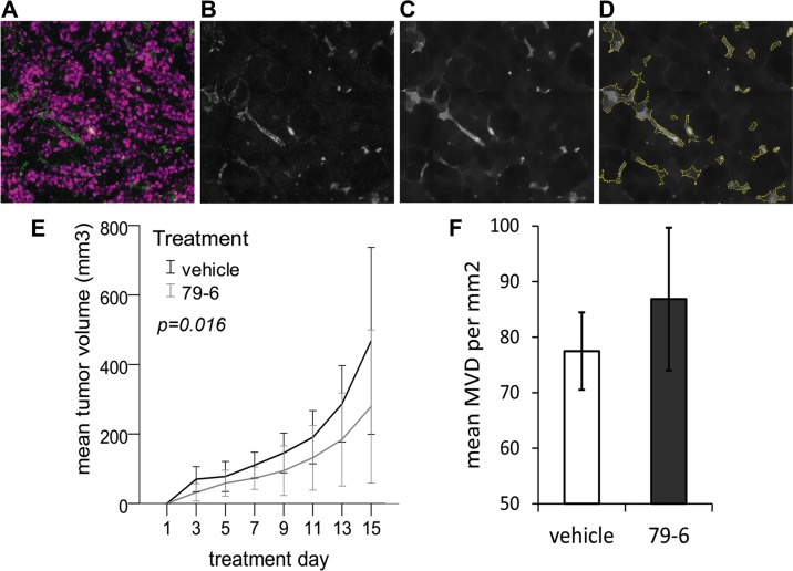
Figure 7. Effect of the Bcl-6 inhibitor 79-6 on HT-29 tumor growth and microvessel density.
BALB/c nude mice received subcutaneous injections of 1 × 10⁷ tumor cells into the right flank and were subsequently treated by intraperitoneal injections of 79-6 (N = 8) or vehicle (N = 8) for 14 consecutive days. Tumor growth (E) was recorded every second day. One day after the last treatment, animals were sacrificed and cryosections of tumors were stained for the endothelial marker CD31. Analysis of microvessel density (MVD) was based on automated tissue scanning by TissueFAXS and an algorithm developed with StrataQuest 5.0 software (TissueGnostics) as exemplified in A–D. Based on fluorescence images (A) of cell nuclei (purple) and endothelial CD31 expression (green), the FITC channel (CD31) was extracted to an 8-bit image, followed by a blurring algorithm (B) after which a morphological mask (C) was applied. (D) Microvessels were identified and quantitated based on kernel radius and background threshold settings which were manually adapted for each batch of scanned images. Results (mean and standard deviation) from three independent analyses are shown in (F).

